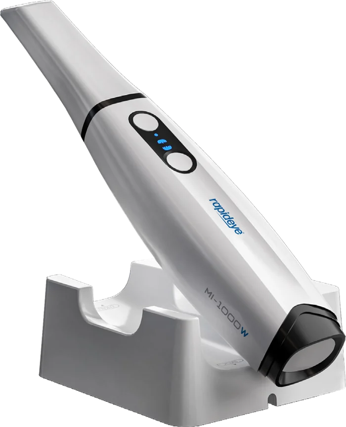
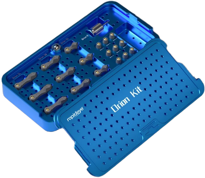
A complete digital workflow with options at every stage: guided surgery, scanners, photogrammetry, libraries and prosthetic components, each made to fit the next.
01Planning
Start with the End in Mind
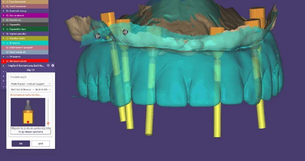
Implant position, soft tissue profile and restorative needs are analysed together before design begins. MODE libraries bring the implant and the M-Guide guided surgery kit into your planning software.
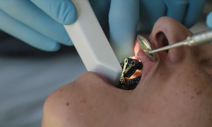
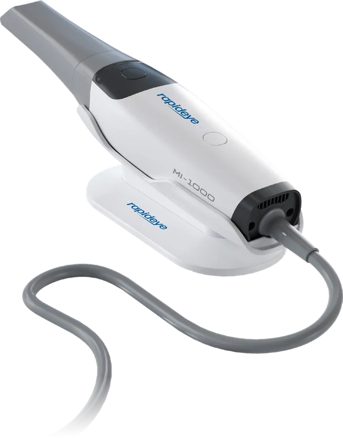
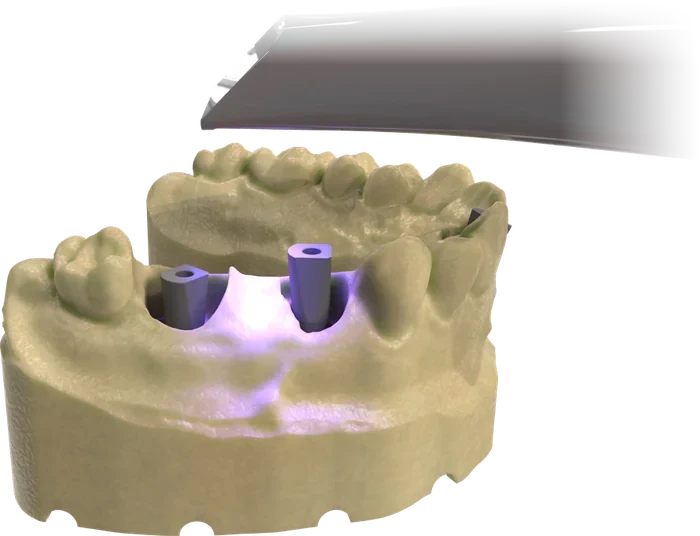
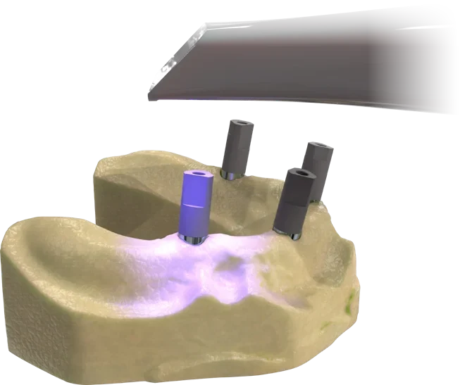
RAPIDEYE intraoral scanners capture the arch, and MODE scanbodies transfer the position and angulation of each implant, from a single implant to an edentulous arch.
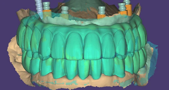
The restoration is designed on the selected MODE component, considering function, occlusal relationships and adjacent teeth. Ti-Base and Multi-Unit libraries are ready for your CAD software.
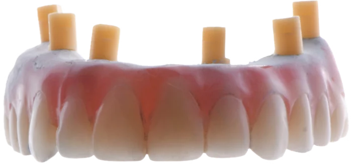
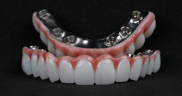
The prosthesis is manufactured from the digital data. Digital analogs position the implants in the model; Ti-Bases, digital copings and bar connections join the restoration to the implant.
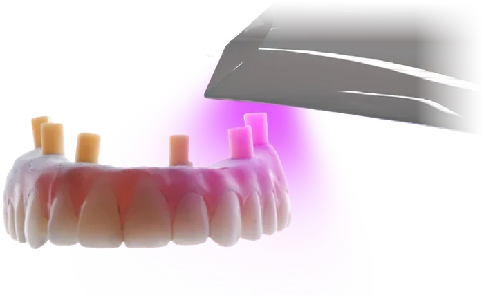
The restoration is placed on the prosthetic components and the connection fit is verified. Occlusion and intraoral adaptation are evaluated, and the treatment is completed.
The same digital workflow scales from one crown to a complete arch. Select a case to see its components and steps.
Ti-Base Abutment Series · Engaged
One Implant, One Crown
Engaged Ti-Bases in Standard, Long and Angled Flex options, with the scanbody and digital analog for the same connection.
Standard Ti-Base4 mm
Long Ti-Base7 mm
Flex Ti-Baseup to 20°
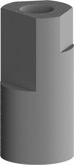
Scanbody
Digital Analog
1Soft Tissue Scanning
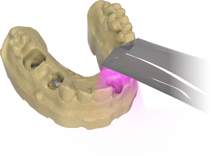
Soft tissue around the implant is digitally scanned to help define the restorative margins. This supports esthetic integration and more accurate planning of the emergence profile.
2Single Implant Scanning
The scanbody is placed on the implant to transfer its position and connection data to the digital environment: the essential data for a single implant case.
3Selection of the Ti-Base
Each case requirement is evaluated to select the ideal component from the Long, Standard or Flex Ti-Base range, for an optimized emergence profile.
4Case Analysis and Planning
Implant position, soft tissue profile and restoration needs are analysed together before digital design, increasing predictability throughout the treatment.
5Digital Crown and Bridge Design
The restoration is designed on the selected prosthetic component, considering function, occlusal relationships and adjacent tooth alignment.
6Clinical Try-In and Initial Evaluation
The restoration is placed on the prosthetic components and the connection fit is verified, before final intraoral adjustment.
7Final Adjustment and Delivery
Occlusion, connection fit and intraoral adaptation are evaluated. After any necessary adjustments, the restoration is finalized.
Ti-Base Non-Engaged · Multi-Unit
Several Implants, One Restoration
Non-engaged Ti-Bases and Multi-Unit abutments for bridges, with scanning components that transfer the position and angulation of every implant.
Non-Engaged Ti-BaseStandard, Long, Flex
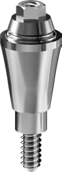
Multi-Unit Abutmentstraight
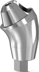
Angled Multi-Unit17° and 30°
Scanbody
Multi-Unit Scanbody
1Soft Tissue Scanning
Soft tissue around the implant is digitally scanned to help define the restorative margins. This supports esthetic integration and more accurate planning of the emergence profile.
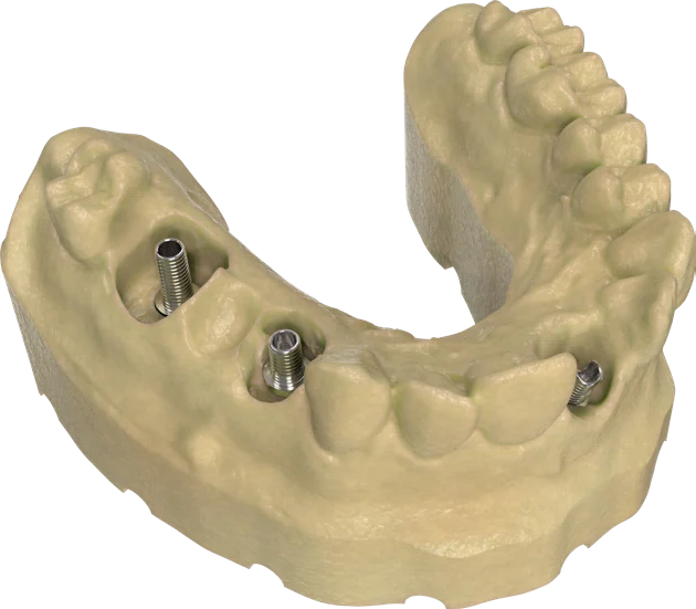
2Scanning in Multi-Implant Cases
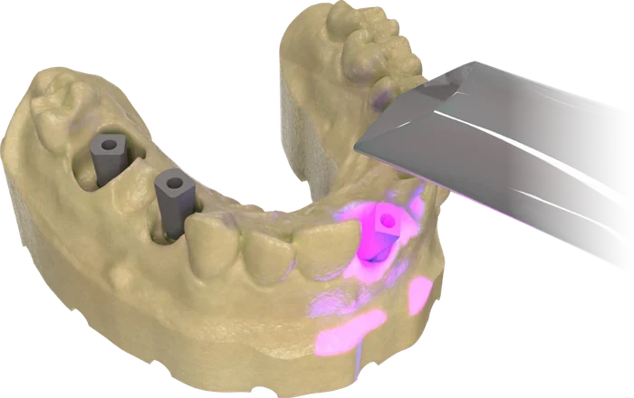
Scanning components transfer the position and angulation of every implant. The resulting data enables final restorations without trials in multi-implant cases.
3Selection of the Ti-Base
Each case requirement is evaluated to select the ideal component from the Long, Standard or Flex Ti-Base range, for an optimized emergence profile.
4Case Analysis and Planning
Implant position, soft tissue profile and restoration needs are analysed together before digital design, increasing predictability throughout the treatment.
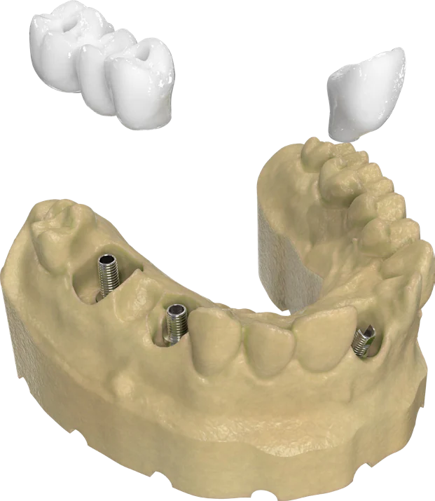
5Digital Crown and Bridge Design
The restoration is designed on the selected prosthetic component, considering function, occlusal relationships and adjacent tooth alignment.
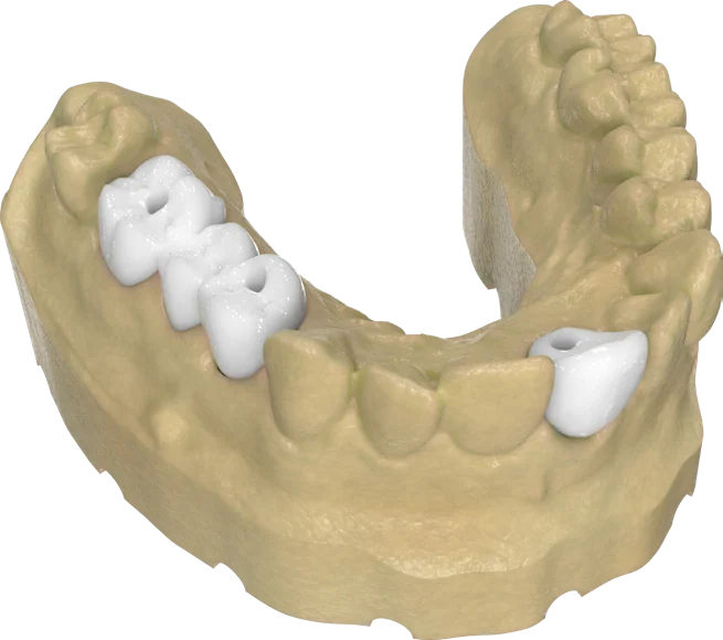
6Clinical Try-In and Initial Evaluation
The restoration is placed on the prosthetic components and the connection fit is verified, before final intraoral adjustment.
7Final Adjustment and Delivery
Occlusion, connection fit and intraoral adaptation are evaluated. After any necessary adjustments, the restoration is finalized.
Multi-Unit Abutment Series
A Full Arch, Planned for Passive Fit
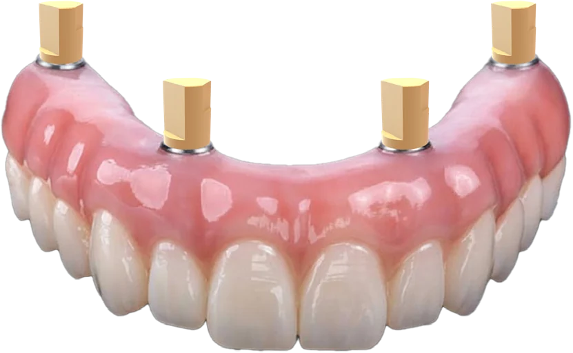
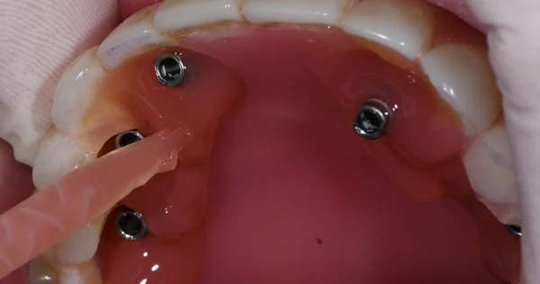
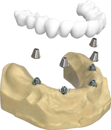
Straight, 17° and 30° Multi-Unit abutments adapt to implant position. The Multi Scanning Kit and Reverse Scanbody are made for edentulous cases.
Multi-Unit Abutmentstraight
Angled Multi-Unit17° and 30°
Multi-Unit Scanbody
Multi Scanning Kit
Reverse Scanbody
Digital Coping
1Case Evaluation and Abutment Selection
Implant positions are evaluated according to the treatment plan, and the appropriate Multi-Unit abutments are selected.
2Soft Tissue Scan
Soft tissue around the implant is digitally captured to support accurate planning of the restorative margins.
3Scanning of Multi-Unit Scanbodies
Scanbodies are placed to transfer the position and angulation data of the implants to the digital environment.
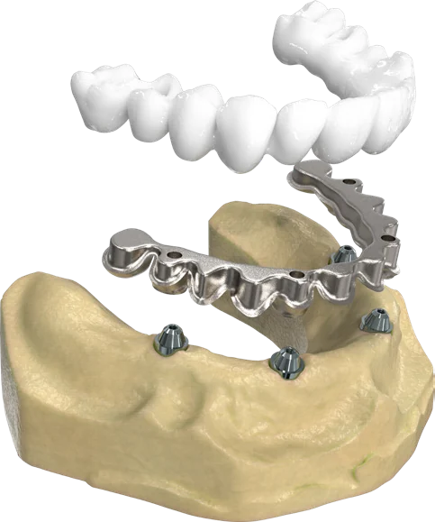
4Coping Connection
Supports passive-fit restorations by providing a stable foundation for monolithic zirconia and temporary restorations.
5I-Bar Connection
An alternative connection option to meet different clinical requirements and support restorative flexibility.
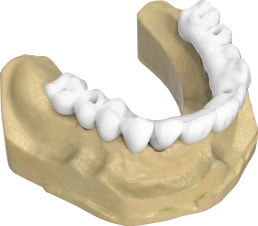
6Prosthetic Seating and Fit
The restoration is seated, and connection integrity and intraoral fit are then evaluated.
Ti-Base Abutment Series
For single-unit and bridge restorations made with CAD/CAM systems. Engaged and non-engaged, for NP and RP platforms.
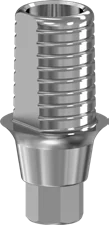
Standard · 4 mm
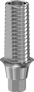
Long · 7 mm
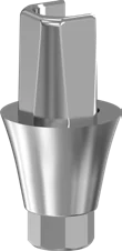
Flex · 4.3 mm
1
Square-Cut Post
A prep-form geometry with flat surfaces for anti-rotational seating and a homogeneous cement gap.
2
Angled Screw Channel
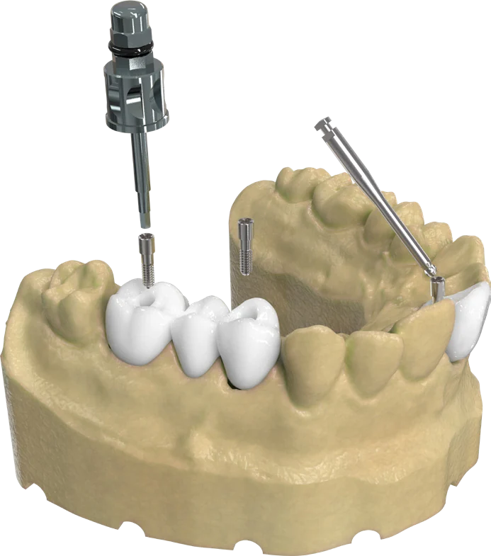
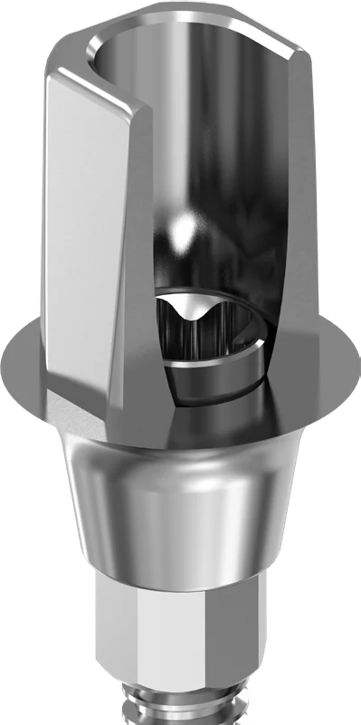
Flex Ti-Base supports angulations of up to 20°. The Flex Screw and Flex Driver are designed for stable torque transfer.
3
Conical Connection
A precise conical connection and a robust connection architecture.
Standard Ti-Base
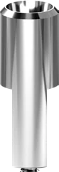
Ideal for restorations with a 4 mm crown connection height. Suitable for single-unit and multi-unit restorations compatible with CAD/CAM systems.
Long Ti-Base New
The 7 mm option is intended for deeply placed implants or restorations requiring greater vertical height.
Flex Ti-Base New
Optimized for angled screw channels of up to 20°, offering improved esthetics and greater flexibility in challenging cases.
Flex Screw New
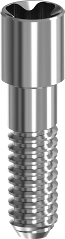
Developed for Ti-Base restorations, with a specialized head design compatible with angled screw channels.
Flex Driver New
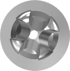
Supports angulations of up to 20° for easier access to angled screw channels, with stable and reliable torque transfer.
Every Ti-Base is available for NP and RP platforms, Engaged and Non-Engaged, in two heights: 0.7 and 2.5 mm (Flex: 1.0 and 2.5 mm). The screw head and driver interface help reduce the risk of slipping or stripping during clinical use.
Multi-Unit Abutment Series
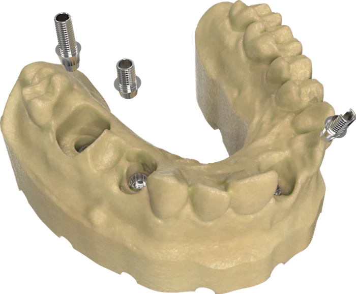
For screw-retained bridge and full-arch restorations, with a transgingival contour optimized for natural gingival anatomy and full compatibility with global CAD/CAM libraries and intraoral scanners.
Straight
17° angled
30° angled
1
Universal Prosthetic Connection
Supports a wide range of screw-retained restorations, with a seamless 360° platform for stability and passive fit.
2
Long Conical Connection
Stable seating and sealing through a 1.44 mm implant-abutment conical contact surface.
3
Octagonal Connection
More indexing options than trilobe, square or hexagonal designs, helping improve passive fit in multi-unit restorations.
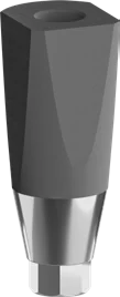
Standard Multi-Unit
NP in heights of 1.5, 2.5 and 3.5 mm. RP in heights of 1.5, 2.5, 3.5 and 4.5 mm.
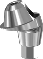
17° Angled Multi-Unit New
NP and RP in heights of 1.5, 2.5 and 3.5 mm.
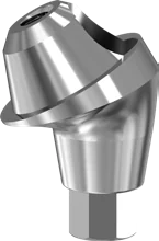
30° Angled Multi-Unit New
NP and RP in heights of 2.5, 3.5 and 4.5 mm.
Multi-Unit Digital Coping
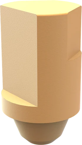
A 4 mm coping that gives monolithic zirconia and temporary restorations a stable foundation.
Gingival height options adapt to different gingival levels, supporting soft tissue management and a controlled emergence profile. The series offers a passive fit tolerance of up to 30° for angled implant placements, with an M1.4 prosthetic screw in a fully enclosed channel.
Full-Arch Precision Scanning Solutions
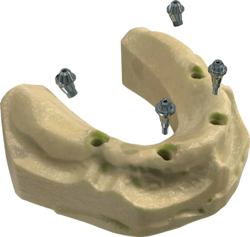
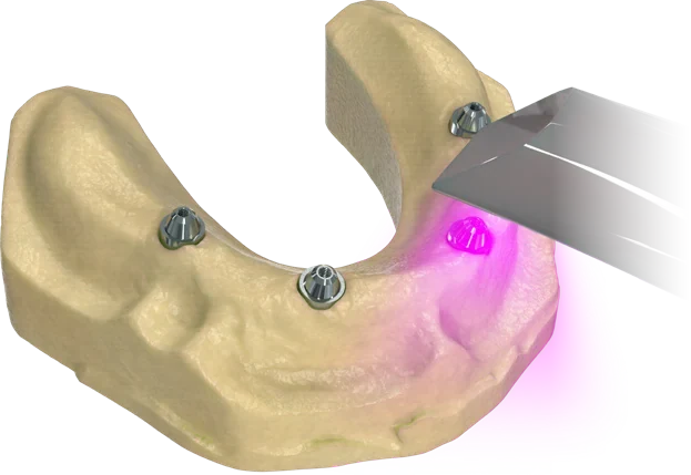
Intraoral scanners rely on reference points in the soft-tissue topography. In edentulous cases, capturing them from soft tissue can slow scanning and increase the risk of cross-arch deviation.
Multi-Unit Scanbody
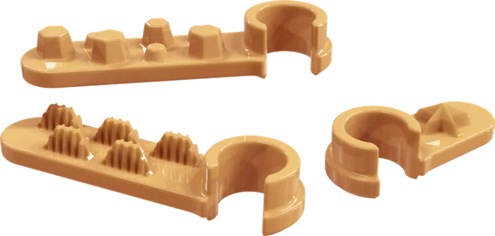
Designed for accurate digital registration of both straight and angled Multi-Unit abutments. It is manufactured from titanium, and its sandblasted surface helps reduce reflections and supports faster digital matching.
Multi Scanning Kit New
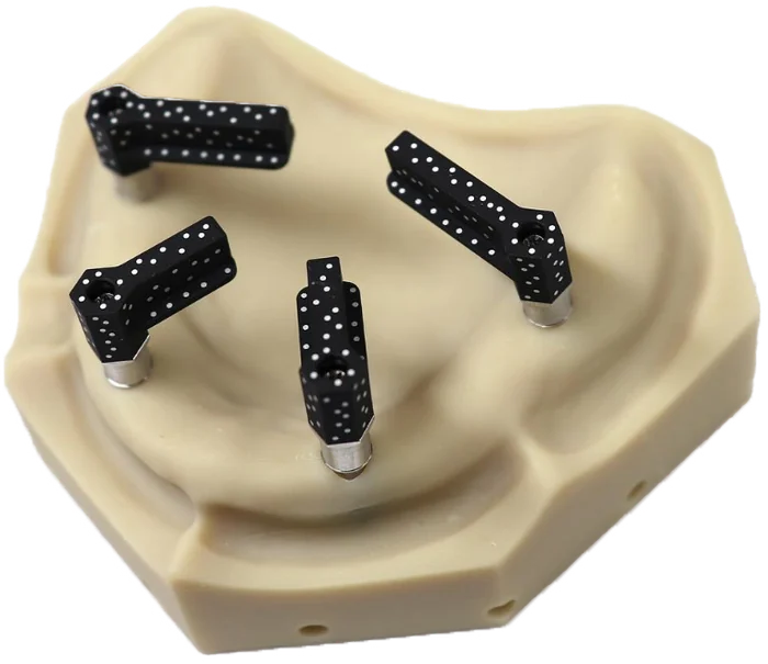
Connected to the Multi-Units, the kit gives the scanner rigid reference points across the arch. Scan efficiency is improved, cross-arch deviation is minimized and excessive overlapping scan data is reduced.
Reverse Scanbody New
Designed for edentulous cases on the reverse scanning principle: implant positions are transferred to the digital environment with high accuracy, directly through the temporary restoration.
Digital Workflow with the Reverse Scanbody
The clinically approved temporary prosthesis becomes the reference for the final one.
1Referencing the Temporary Prosthesis
A temporary prosthesis with clinically verified function and fit is used as a reference: the foundation for accurate and reliable recording of implant positions.
2Connecting the Reverse Scanbodies
Reverse Scanbodies are attached to the temporary prosthesis to read the implant positions, so implant coordinates are transferred with high accuracy.
3Intraoral Scanning in a Clinical Setting
With the scanbodies attached, the temporary prosthesis is transferred to the digital environment with an intraoral scanner, reducing the number of patient visits.
4Digital Matching
Through the scan data, the Reverse Scanbodies are matched within the software to clarify implant positions for restorative planning.
5Digital Design of the Final Prosthesis
The final restoration is designed using the approved temporary prosthesis as a reference, preserving functional and esthetic data while accelerating design.
6Production and Passive Fit
The final prosthesis is manufactured from the data obtained through the Reverse Scanbody protocol, supporting passive fit in clinical application.
Photogrammetry-Assisted Scanning Systems
The MODE digital workflow operates in full compatibility with photogrammetry-based intraoral and extraoral scanning technologies.
Intraoral
Photogrammetry compatibility for precision full-arch scanning, inside the mouth.
Compatibility with global devices and library support
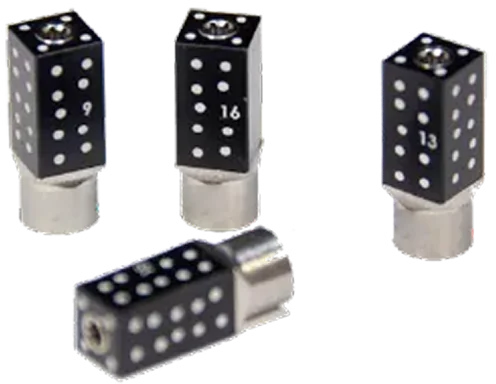
Compatibility with Multi-Unit photogrammetry scan bodies
Guaranteed precision results within seconds
Eliminates RPT risks
Extraoral
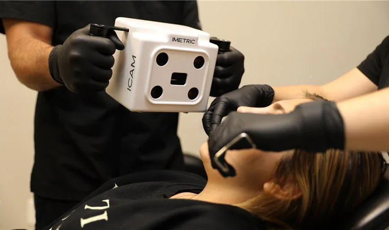
Photogrammetry compatibility for precision full-arch scanning, with an extraoral camera.
Compatibility with global devices and library support
Compatibility with Multi-Unit photogrammetry scan bodies
Guaranteed precision results within seconds
Eliminates RPT risks
Compatible with global extraoral precision scanning systems such as Imetric, Oxo and others.
For full-arch photogrammetry within the ecosystem, see the RAPIDEYE Orion Kit. Imetric®, Oxo® and other trademarks mentioned on this page are the property of their respective owners.
Compatible with Your Digital Workflow
Alongside the RAPIDEYE scanners in the ecosystem, MODE components and libraries work with common scanners, planning and design software and stability measurement devices.
Kişisel verileriniz; veri sorumlusu olarak Firma Adı (“ŞİRKET” veya Firma Adı” olarak adlandırılacaktır.) tarafından işletilen (www.alanadi.com) internet sitesini ziyaret edenlerin gizliliğini korumak Kurumumuzun önde gelen ilkelerindendir. Bu Çerez Kullanımı Politikası (“Politika”), tüm web sitesi ziyaretçilerimize ve kullanıcılarımıza hangi tür çerezlerin hangi koşullarda kullanıldığını açıklamaktadır.
Çerezler, bilgisayarınız ya da mobil cihazınız üzerinden ziyaret ettiğiniz internet siteleri tarafından cihazınıza veya ağ sunucusuna depolanan küçük metin dosyalarıdır.
Genellikle ziyaret ettiğiniz internet sitesini kullanmanız sırasında size kişiselleştirilmiş bir deneyim sunmak, sunulan hizmetleri geliştirmek ve deneyiminizi iyileştirmek için kullanılır ve bir internet sitesinde gezinirken kullanım kolaylığına katkıda bulunabilir. Çerez kullanılmasını tercih etmezseniz tarayıcınızın ayarlarından Çerezleri silebilir ya da engelleyebilirsiniz. Ancak bunun internet sitemizi kullanımınızı etkileyebileceğini hatırlatmak isteriz. Tarayıcınızdan Çerez ayarlarınızı değiştirmediğiniz sürece bu sitede çerez kullanımını kabul ettiğinizi varsayacağız.
1. ÇEREZLERDE HANGİ TÜR VERİLER İŞLENİR?
İnternet sitelerinde yer alan çerezlerde, türüne bağlı olarak, siteyi ziyaret ettiğiniz cihazdaki tarama ve kullanım tercihlerinize ilişkin veriler toplanmaktadır. Bu veriler, eriştiğiniz sayfalar, incelediğiniz hizmet ve ürünler, tercih ettiğiniz dil seçeneği ve diğer tercihlerinize dair bilgileri kapsamaktadır.
2. ÇEREZ NEDİR ve KULLANIM AMAÇLARI NELERDİR?
Çerezler, ziyaret ettiğiniz internet siteleri tarafından tarayıcılar aracılığıyla cihazınıza veya ağ sunucusuna depolanan küçük metin dosyalarıdır. Sitede tercih ettiğiniz dil ve diğer ayarları içeren bu küçük metin dosyaları, siteye bir sonraki ziyaretinizde tercihlerinizin hatırlanmasına ve sitedeki deneyiminizi iyileştirmek için hizmetlerimizde geliştirmeler yapmamıza yardımcı olur. Böylece bir sonraki ziyaretinizde daha iyi ve kişiselleştirilmiş bir kullanım deneyimi yaşayabilirsiniz.
İnternet Sitemizde çerez kullanılmasının başlıca amaçları aşağıda sıralanmaktadır:
İnternet sitesinin işlevselliğini ve performansını arttırmak yoluyla sizlere sunulan hizmetleri geliştirmek,
İnternet Sitesini iyileştirmek ve İnternet Sitesi üzerinden yeni özellikler sunmak ve sunulan özellikleri sizlerin tercihlerine göre kişiselleştirmek;
İnternet Sitesinin, sizin ve Kurum’un hukuki ve ticari güvenliğinin teminini sağlamak, Site üzerinden sahte işlemlerin gerçekleştirilmesini önlemek;
5651 sayılı Internet Ortamında Yapılan Yayınların Düzenlenmesi ve Bu Yayınlar Yoluyla İşlenen Suçlarla Mücadele Edilmesi Hakkında Kanun ve Internet Ortamında Yapılan Yayınların Düzenlenmesine Dair Usul ve Esaslar Hakkında Yönetmelik’ten kaynaklananlar başta olmak üzere, kanuni ve sözleşmesel yükümlülüklerini yerine getirmek.
3.İNTERNET SİTEMİZDE KULLANILAN ÇEREZ TÜRLERİ
3.1.Oturum Çerezleri
Oturum çerezlerini ziyaretinizi süresince internet sitesinin düzgün bir şekilde çalışmasının teminini sağlamaktadır. Sitelerimizin ve sizin, ziyaretinizde güvenliğini, sürekliliğini sağlamak gibi amaçlarla kullanılırlar. Oturum çerezleri geçici çerezlerdir, siz tarayıcınızı kapatıp sitemize tekrar geldiğinizde silinir, kalıcı değillerdir.
3.2.Kalıcı Çerezler
Bu tür çerezler tercihlerinizi hatırlamak için kullanılır ve tarayıcılar vasıtasıyla cihazınızda depolanır Kalıcı çerezler, sitemizi ziyaret ettiğiniz tarayıcınızı kapattıktan veya bilgisayarınızı yeniden başlattıktan sonra bile saklı kalır. Tarayıcınızın ayarlarından silinene kadar bu çerezler tarayıcınızın alt klasörlerinde tutulurlar.
Kalıcı çerezlerin bazı türleri; İnternet Sitesini kullanım amacınız gibi hususlar göz önünde bulundurarak sizlere özel öneriler sunulması için kullanılabilmektedir.
Kalıcı çerezler sayesinde İnternet Sitemizi aynı cihazla tekrardan ziyaret etmeniz durumunda, cihazınızda İnternet Sitemiz tarafından oluşturulmuş bir çerez olup olmadığı kontrol edilir ve var ise, sizin siteyi daha önce ziyaret ettiğiniz anlaşılır ve size iletilecek içerik bu doğrultuda belirlenir ve böylelikle sizlere daha iyi bir hizmet sunulur.
3.3.Zorunlu/Teknik Çerezler
Ziyaret ettiğiniz internet sitesinin düzgün şekilde çalışabilmesi için zorunlu çerezlerdir. Bu tür çerezlerin amacı, sitenin çalışmasını sağlamak yoluyla gerekli hizmet sunmaktır. Örneğin, internet sitesinin güvenli bölümlerine erişmeye, özelliklerini kullanabilmeye, üzerinde gezinti yapabilmeye olanak verir.
3.4.Analitik Çerezler
İnternet sitesinin kullanım şekli, ziyaret sıklığı ve sayısı, hakkında bilgi toplayan ve ziyaretçilerin siteye nasıl geçtiğini gösterirler. Bu tür çerezlerin kullanım amacı, sitenin işleyiş biçimini iyileştirerek performans arttırmak ve genel eğilim yönünü belirlemektir. Ziyaretçi kimliklerinin tespitini sağlayabilecek verileri içermezler. Örneğin, gösterilen hata mesajı sayısı veya en çok ziyaret edilen sayfaları gösterirler.
3.5.İşlevsel/Fonksiyonel Çerezler
Ziyaretçinin site içerisinde yaptığı seçimleri kaydederek bir sonraki ziyarette hatırlar. Bu tür çerezlerin amacı ziyaretçilere kullanım kolaylığı sağlamaktır. Örneğin, site kullanıcısının ziyaret ettiği her bir sayfada kullanıcı şifresini tekrar girmesini önler.
3.6. Hedefleme/Reklam Çerezleri
Ziyaretçilere sunulan reklamların etkinliğinin ölçülmesi ve reklamların kaç kere görüntülendiğinin hesaplanmasını sağlarlar. Bu tür çerezlerin amacı, ziyaretçilerin ilgi alanlarına özelleştirilmiş reklamların sunulmasıdır.
Aynı şekilde, ziyaretçilerin gezinmelerine özel olarak ilgi alanlarının tespit edilmesini ve uygun içeriklerin sunulmasını sağlarlar. Örneğin, ziyaretçiye gösterilen reklamın kısa süre içinde tekrar gösterilmesini engeller.
4.ÇEREZ TERCİHLERİ NASIL YÖNETİLİR?
Çerezlerin kullanımına ilişkin tercihlerinizi değiştirmek ya da çerezleri engellemek veya silmek için tarayıcınızın ayarlarını değiştirmeniz yeterlidir.
Birçok tarayıcı çerezleri kontrol edebilmeniz için size çerezleri kabul etme veya reddetme, yalnızca belirli türdeki çerezleri kabul etme ya da bir internet sitesinin cihazınıza çerez depolamayı talep ettiğinde tarayıcı tarafından uyarılma seçeneği sunar.
Aynı zamanda, daha önce tarayıcınıza kaydedilmiş çerezlerin silinmesi de mümkündür.
Çerezleri devre dışı bırakır veya reddederseniz, bazı tercihleri manuel olarak ayarlamanız gerekebilir, hesabınızı tanıyamayacağımız ve ilişkilendiremeyeceğimiz için internet sitesindeki bazı özellikler ve hizmetler düzgün çalışmayabilir. Tarayıcınızın ayarlarını aşağıdaki tablodan ilgili link’e tıklayarak değiştirebilirsiniz.
İnternet Sitesi Gizlilik Politikası …./…./…. tarihlidir. Politika’nın tümünün veya belirli maddelerinin yenilenmesi durumunda Politika’nın yürürlük tarihi güncellenecektir. Gizlilik Politikası Kurum’un internet sitesinde (www.alanadi.com) yayımlanır ve kişisel veri sahiplerinin talebi üzerine ilgili kişilerin erişimine sunulur.
Firma Adı
Adres: Mahalle Adı Sokak Adı. No: 1/A, 34444 İlçe Adı/İl Adı
Telefon: +90 216 555 55 55
E – Posta: mail@alanadi.com
Web Adresi: www.alanadi.com
? Cookie Preferences
We use cookies to enhance your experience
We use essential cookies for site functionality and optional cookies to improve your experience, analyze site usage, and assist in our marketing efforts.





 Flex Screw New
 Flex Driver New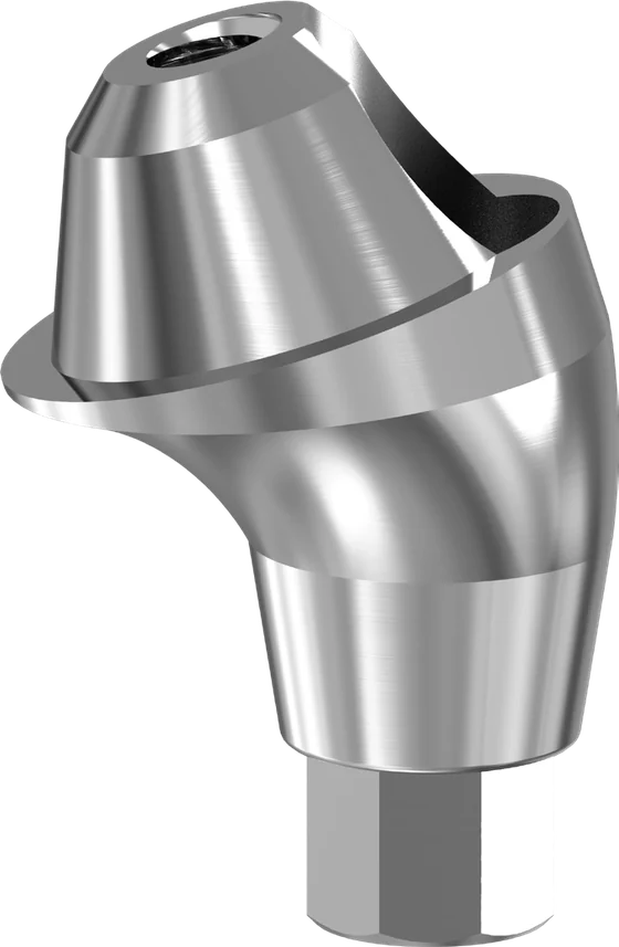
 17° Angled Multi-Unit New
 30° Angled Multi-Unit New